The Huber Lab uses the social amoeba Dictyostelium discoideum as a model system for studying fundamental cellular and developmental processes. Our current research is funded by a NSERC Discovery Grant and a CIHR Project Grant. Our NSERC-funded work is using Dictyostelium as a model system to explore the conserved roles of CLN proteins in regulating processes mediated by the endomembrane system. Our CIHR-funded work is using Dictyostelium and human cell models to study the function of CLN5 with an aim towards understanding how mutations in CLN5 cause Batten disease (neuronal ceroid lipofuscinosis) in humans. The Huber Lab is also interested in using Dictyostelium as a model system for studying the structure and function of the extracellular matrix.
Click on the links below for more information:
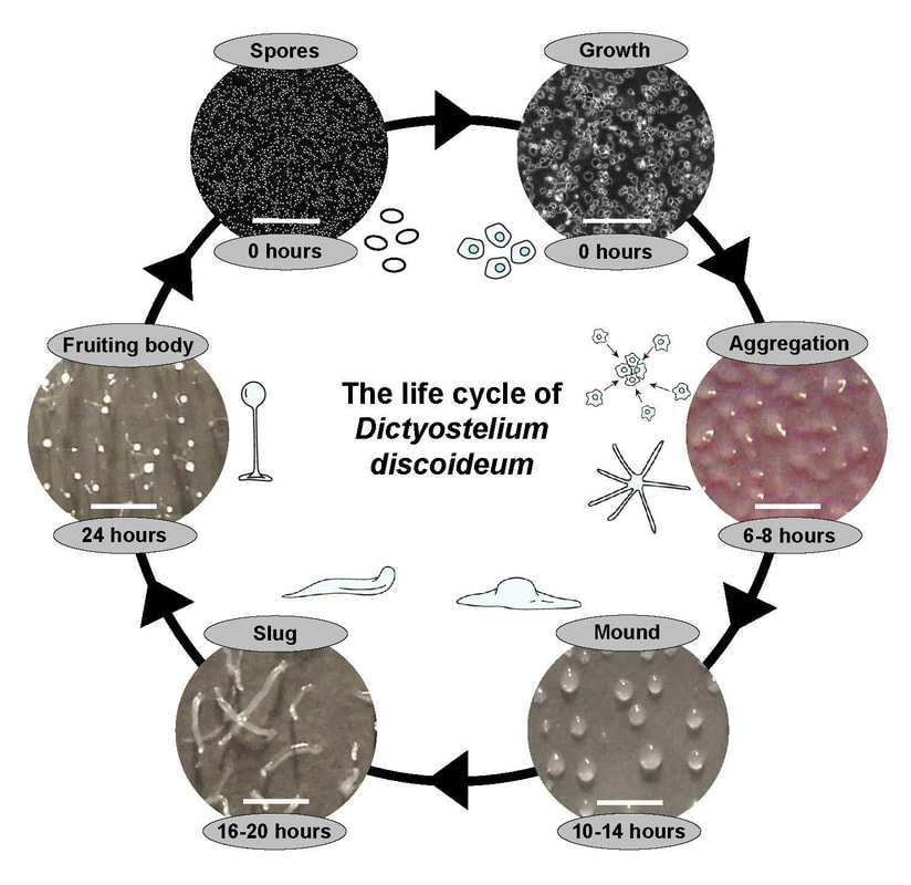
Dictyostelium is a fascinating eukaryotic soil microbe that has long served as a model organism for cell and developmental biology. Inexpensive, easy to culture, and genetically tractable, Dictyostelium undergoes a 24h life cycle comprised of both single-cell and multicellular phases.
|
|
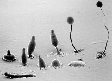
The life cycle of Dictyostelium. During growth, haploid cells feed on bacteria. Upon starvation, cells undergo chemotactic aggregation towards cAMP to form a multicellular mound. The mound then forms a finger, which falls on the surface to generate a motile pseudoplasmodium, also known as a slug. During culmination, terminal differentiation of pre-stalk and pre-spore cells forms a fruiting body composed of a mass of viable spores supported atop a slender stalk. When a food source becomes available, the spores germinate allowing the cells to restart the life cycle. Figure on left taken from Huber et al., 2022. Figure on right Copyright M.J. Grimson & R.L. Blanton, Biological Sciences Electron Microscopy Laboratory, Texas Tech University.
The Dictyostelium genome is haploid and encodes many homologs of genes linked to human disease. The genetic tractability of the organism allows researchers to introduce one or multiple gene deletions with relative ease using homologous recombination or CRISPR/Cas9-mediated targeting. Finally, development occurs in a much shorter time frame in Dictyostelium compared to other organisms, which allows researchers to rapidly screen Dictyostelium mutant cell lines for developmental phenotypes.
The article below provides a great review on how Dictyostelium is used as a model system for cell and developmental biology:
Mathavarajah S+, Flores A+, Huber RJ. (2017). Dictyostelium discoideum: A model system for cell and developmental biology. Current Protocols Essential Laboratory Techniques 15, 14.1.1-14.1.19. doi:10.1002/cpet.15
http://onlinelibrary.wiley.com/doi/10.1002/cpet.15/full
Undergraduate and graduate students interested in contributing to this work are encouraged to contact Dr. Huber with a statement of interest, updated CV, and recent unofficial transcript. Full funding may be available for students who qualify.
The Huber Lab would like to thank the following agencies (past and present) for funding this work
|
|

|

|
|
|
|
|
